


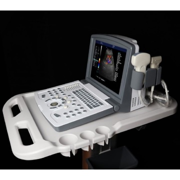
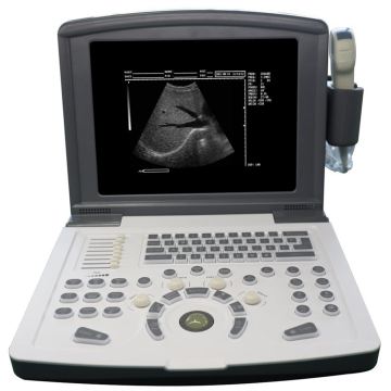
Portable Color Doppler Ultrasound Machine for Small Organ
$2100-3500 /Set/Sets
| Payment Type: | T/T |
| Incoterm: | FOB |
| Min. Order: | 5 Set/Sets |
| Transportation: | Ocean,Land,Express,Air |
| Port: | Shenzhen,Ningbo,Shanghai |

$2100-3500 /Set/Sets
| Payment Type: | T/T |
| Incoterm: | FOB |
| Min. Order: | 5 Set/Sets |
| Transportation: | Ocean,Land,Express,Air |
| Port: | Shenzhen,Ningbo,Shanghai |
Model No.: MDK-680
Brand: United Ultrasound
Place Of Origin: China
Kind Of Power: Electricity
Warranty Service: 1 Year
After-sales Service: Free Spare Parts, Online Technical Support
Material: Metal, Plastic
Medical Device Classification: Class Ii
Brand: United Ultrasound
Monitor Size: 12 inch
Gray Scale: 256 levels
Depth: ≥300mm
Channels: 32
Operating System: Windows 7
Color: black
| Selling Units | : | Set/Sets |
| Package Type | : | Carton |
Portable Color Doppler Ultrasound Machine for Small Organ(MDK-680)
In small organs, such as thyroid, mammary gland and eyeball, color ultrasound has obvious diagnostic accuracy.
From a certain point of view, the 10MHz probe has been more clear than the ordinary black and white Doppler over 5MHz, and the probe is much clearer. The diagnosis and differential diagnosis of thyroid lesions are mainly based on the internal blood supply of the thyroid, among which the hyperthyroidism image is the most typical and specific.
Simple goiter showed no significant change compared with normal thyroid blood flow. Subacute thyroiditis, Hashimoto's thyroiditis between the two, can be distinguished, and through nodules and peripheral blood flow situation can be well distinguished nodular goiter, thyroid adenoma and thyroid cancer, so it is recommended that the thyroid diagnosis is not clear, the patient has a certain economic ability can do color ultrasound further diagnosis.
Breast color ultrasound is mainly used in the differential diagnosis of mammary fibroma and breast cancer, and the eyeball mainly has a better value in the diagnosis of eyeball vascular diseases.
Our products:
Black And White Ultrasound Scanner
Color Doppler Ultrasound Scanner
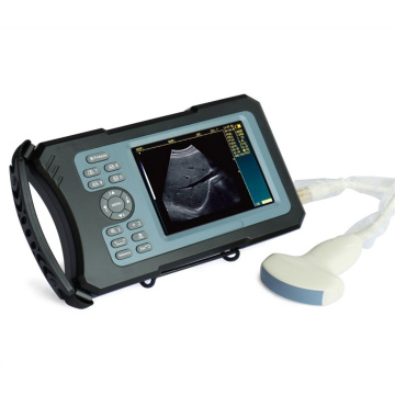
Veterinary Ultrasound Scanner

No. Item Index <1> Depth ≥300mm <2> Lateral Resolution ≤1mm (Depth≤80mm) ≤2mm (80< Depth≤130mm) <3> Axial Resolution ≤1mm (Depth≤80mm) ≤2mm (80< Depth≤130mm) <4> Blind Area ≤5 mm <5> Geometry Position Precision horizontal≤5% vertical≤5% <6> Language English/Chinese <7> Channels 32 <8> Displayer 12” LED <9> External Display PAL, VGA, USB <10> Gray Scale 256 levels <11> Voltage AC220V ±10% <12> Operating System Windows 7 <13> Scanning Mode B, B/B, 4B, B/M, M, B+C, B+D, B+C+D, PDI, CF, PW <14> Probe Probe sockets: 2 Probe frequency: 2.0MHz ~ 10.0MHz, 8-step frequency conversion <15> Adjustment parameters of color blood flow image Doppler frequency, sampling frame position and size, baseline, color gain, deflection angle, wall filtering, cumulative times, etc <16> Signal processing With dynamic filtering and quadrature demodulation With total gain adjustment Gain adjustment: 8-segment TGC The total gain of Type B, Type C and Type D can be adjusted respectively B/W image gain and color blood flow gain are adjustable respectively <17> Doppler Doppler baseline adjustment level 6 Pulse repetition frequency can be adjusted separately: CFM PWD With D linear speed regulation <18> Digital beam forming Continuous dynamic focusing of digital beam forming image Full range dynamic aperture of image Dynamic tracing of the whole image Weighted Sum of Image Whole Process Receiving Delay Support half step scanning and ± 10 ° linear receiving deflection angle Multi beam parallel processing technology <19> Basic measurement and calculation function Basic measurement in mode B: distance, angle, perimeter and area, volume, stenosis rate, histogram, cross-section Basic measurement of M-mode: heart rate, time, distance and speed Doppler measurement: time, heart rate, speed, acceleration


Privacy statement: Your privacy is very important to Us. Our company promises not to disclose your personal information to any external company with out your explicit permission.
Fill in more information so that we can get in touch with you faster
Privacy statement: Your privacy is very important to Us. Our company promises not to disclose your personal information to any external company with out your explicit permission.


